In the past, several hours of micro-CT scans were performed to image rodent vessels after sacrifice, which either infused the blood vessels with radiopaque polymers or shocked the animals infused with contrast media. In in vitro studies, we do not need to reduce the dose of x-rays, and the scan time does not need to be affected by the anesthetic effect. Therefore, the SNR (signal-to-noise ratio) can be maximized by long-time high-energy scanning, thus obtaining sub-micron level. Resolution. Advances in X-ray detector technology have enabled micro-CT to achieve faster read times, larger pixel matrices, and higher X-ray sensitivity and higher SNR. For example, Hitachi Aloka's multi-row quantitative small animal CT LCT200 uses a multi-row high-sensitivity CdTe semiconductor detector. Unlike previous scintillation radiation detectors, this detector does not have a complicated radio signal conversion process, but instead uses X. The ray is directly converted into an electrical signal for analytical imaging, which reduces image blur caused by light scattering during the conversion process, and scans quickly, and high-quality and high-contrast images with high contrast can be obtained. This technological advancement, combined with the instrument's breath scan and heartbeat scan function, eliminates motion artifacts during the scan, while avoiding the anxieties caused by prolonged anesthesia, reducing the radiation dose and reducing the tradition of angiogenesis research. The use of contrast agents.
Kiessling et al. and Schambach et al. have reported high-resolution vascular 3D imaging of living animals, respectively, in which they performed high-resolution imaging of blood vessels and cerebrovascular vessels at the tumor site in mice. In addition, the pathological features of some blood vessels can be detected without the use of contrast agents. For example, Persy et al. evaluated the degree of arterial calcification in rats with long-term renal failure and concluded the degree and development of renal failure in these animals. The study did not use contrast agents.
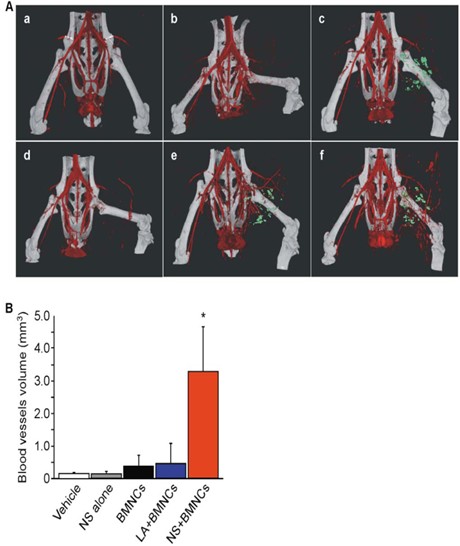
Figure 1. Effect of different treatments on angiogenesis in hind limb ischemia model mice by LCT200 (from PLOS ONE April 2012 | Volume 7 | Issue 4 | e35199)

Figure 2. Imaging results of intracranial blood vessels in mice using conventional contrast agents (Iomerone 300, Bracco Altana)
Fresh Carrot
Carrot (scientific name: Daucus Carota var. sativa Hoffm.) is a variety of wild carrot in Umbelliferae and carrot genus. It is an annual or biennial herb. The root is strong, long conical, orange red or yellow. Stem erect, up to 90 cm high, much branched. Leaf blade long stalked, pinnately compound, lobes linear or lanceolate, apex acute; Petiole base enlarged to form leaf sheath. Compound umbel; Peduncle hispid; The involucral bracts are mostly leafy, and the umbrella spokes at the outer edge of the fruiting stage bend inward; Flowers usually white, sometimes reddish; Petiole unequal. The fruit is conical, with white thorns on the edges. It blooms in April.
Carotene is the main source of vitamin A, which can promote growth, prevent bacterial infection, and protect epidermal tissue, respiratory tract, digestive tract, urinary system and other epithelial cells; Carrots contain a kind of quercetin, which can increase coronary blood flow, promote adrenaline synthesis, reduce blood pressure and reduce inflammation. The oil content of carrot seeds is up to 13%, which can drive Ascaris lumbricoides and cure dysentery for a long time. Carrot leaves can prevent chickenpox and acute jaundice hepatitis. Long term drinking carrot juice can prevent night blindness and dry eye disease, enrich skin, flatten wrinkles, eliminate spots and keep hair fit. Especially for smokers, eating carrot every day has a better effect on preventing lung cancer.
Fresh Carrot,Storing Carrots,Storing Fresh Carrots,Keeping Carrots Fresh
Tianshui Wenpu International Trade Co.,Ltd , https://www.wenpugroup.com